Beranda
/ Anatomy Of Ribs - 3d Rendered Anatomy Illustration Of The Dog Skeletal Anatomy Ribs Stock Photo Alamy / The rib cage is often simplified as an oval shape.
Anatomy Of Ribs - 3d Rendered Anatomy Illustration Of The Dog Skeletal Anatomy Ribs Stock Photo Alamy / The rib cage is often simplified as an oval shape.
Insurance Gas/Electricity Loans Mortgage Attorney Lawyer Donate Conference Call Degree Credit Treatment Software Classes Recovery Trading Rehab Hosting Transfer Cord Blood Claim compensation mesothelioma mesothelioma attorney Houston car accident lawyer moreno valley can you sue a doctor for wrong diagnosis doctorate in security top online doctoral programs in business educational leadership doctoral programs online car accident doctor atlanta car accident doctor atlanta accident attorney rancho Cucamonga truck accident attorney san Antonio ONLINE BUSINESS DEGREE PROGRAMS ACCREDITED online accredited psychology degree masters degree in human resources online public administration masters degree online bitcoin merchant account bitcoin merchant services compare car insurance auto insurance troy mi seo explanation digital marketing degree floridaseo company fitness showrooms stamfordct how to work more efficiently seowordpress tips meaning of seo what is an seo what does an seo do what seo stands for best seotips google seo advice seo steps, The secure cloud-based platform for smart service delivery. Safelink is used by legal, professional and financial services to protect sensitive information, accelerate business processes and increase productivity. Use Safelink to collaborate securely with clients, colleagues and external parties. Safelink has a menu of workspace types with advanced features for dispute resolution, running deals and customised client portal creation. All data is encrypted (at rest and in transit and you retain your own encryption keys. Our titan security framework ensures your data is secure and you even have the option to choose your own data location from Channel Islands, London (UK), Dublin (EU), Australia.
Anatomy Of Ribs - 3d Rendered Anatomy Illustration Of The Dog Skeletal Anatomy Ribs Stock Photo Alamy / The rib cage is often simplified as an oval shape.. They are twelve in number on either side; True, false, floating, typical, and atypical ribs. The first seven ribs progressively increase in length, the lower five ribs then begin to decrease in length. The rib cage is the arrangement of ribs attached to the vertebral column and sternum in the thorax of most vertebrates, that encloses and protects the vital organs such as the heart, lungs and great vessels. The rib cage is often simplified as an oval shape.
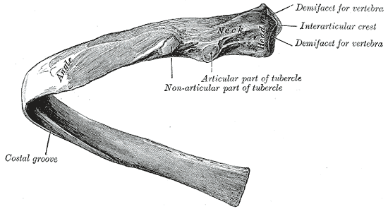
For more anatomy content please follow us and visit our website: The tubercle is a bony prominence. For a gesture drawing, that's good enough. The first seven sets of ribs, known as true ribs also known as vertebrosternal ribs, are directly articulate with the vertebral column posteriorly and terminate anteriorly as costal cartilage. A bicipital rib is seen in relation to the first thoracic rib.
Ribs Physiopedia from www.physio-pedia.com The ribs are located in the chest (thoracic. Ribs anatomy true ribs, false ribs, and floating ribs. The first seven ribs progressively increase in length, the lower five ribs then begin to decrease in length. It appears to be the result of the fusion of two ribs, either of a cervical and first thoracic or of the first two thoracic ribs. They also serve as an attachment point for many muscles and are active during respiration. The rib cage is collectively made up of long, curved. The rib cage is a bony structure found in the chest (thoracic cavity). Affiliation 1 section of thoracic and.
Costae) are long, flat, curved bones that form the rib cage.
They articulate with the vertebral column posteriorly, and terminate anteriorly as cartilage (known as costal cartilage). The heart is a muscle at the center of your circulatory system. It appears to be the result of the fusion of two ribs, either of a cervical and first thoracic or of the first two thoracic ribs. Related posts of abdominal diagram with ribs abdominal anatomy muscles. They also serve as an attachment point for many muscles and are active during respiration. The front view of the thorax. Head, neck, body or shaft, tubercle, and angle. Costae) are long, flat, curved bones that form the rib cage. These videos are for educational purpose only for the medical students like mbb. Anatomynote.com found heart, lung, diaphragm and ribs location from plenty of anatomical pictures on the internet. There are twelve (12) pairs of ribs and all articulate posteriorly with the thoracic vertebrae. The first seven sets of ribs, known as true ribs also known as vertebrosternal ribs, are directly articulate with the vertebral column posteriorly and terminate anteriorly as costal cartilage. Ribs anatomy true ribs, false ribs, and floating ribs.
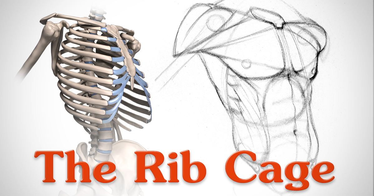
With each succeeding rib, from the first, or uppermost, the curvature of the rib cage becomes more open. Ribs the ribs partially enclose and protect the chest cavity, where many vital organs (including the heart and the lungs) are located. True, false, floating, typical, and atypical ribs. An individual rib has five parts: The rib cage is a bony structure found in the chest (thoracic cavity).
How Many Ribs Do Humans Have Men Women And Anatomy from i0.wp.com Authors geoffrey m graeber 1 , muhammad nazim. Abdominal anatomy muscles 12 photos of the abdominal anatomy muscles abdominal muscles anatomy and function, abdominal muscles anatomy diagram, abdominal muscles cross sectional anatomy, deep abdominal muscles anatomy, lateral abdominal muscles anatomy, human anatomy, abdominal muscles anatomy and function, abdominal. The anatomy of the ribs and the sternum and their relationship to chest wall structure and function thorac surg clin. The anatomy of the human ribs is made up of 24 ribs which are parted in 12 pairs (each on the left and right side of the chest wall), with the sternum, metasternum (the xiphoid process), and the costal cartilages all situated at the anterior of the chest wall, followed by the thoracic vertebrae on the posterior of the chest wall. It has clear front, side, and back planes. The right upper quadrant of the abdomen includes the pancreas, right kidney, gallbladder, liver, and intestines. Your heart sits in the middle of your chest, to the left. The head of each rib is wedge shaped and has two articular facets, which are separated by a wedge of bone, known.
The right upper quadrant of the abdomen includes the pancreas, right kidney, gallbladder, liver, and intestines.
There are 3 types of ribs: For a gesture drawing, that's good enough. They are twelve in number on either side; Abdominal anatomy muscles 12 photos of the abdominal anatomy muscles abdominal muscles anatomy and function, abdominal muscles anatomy diagram, abdominal muscles cross sectional anatomy, deep abdominal muscles anatomy, lateral abdominal muscles anatomy, human anatomy, abdominal muscles anatomy and function, abdominal. Affiliation 1 section of thoracic and. They also serve as an attachment point for many muscles and are active during respiration. Ribs are highly vascular and trabecular with a thin outer layer of compact bone. The anatomy of a floating rib anatomy. The rib cage surrounds the lungs and the heart, serving as an important means of bony protection for these vital organs.in total, the rib cage consists of the 12 thoracic vertebrae and the 24 ribs, in addition to the sternum. Costae) are long, flat, curved bones that form the rib cage. In this video, we explore:1) the anatomy of the sternum2) the anatomy and differences between the three classes of ribs3) the anatomy and differences between. The anatomy of the human ribs is made up of 24 ribs which are parted in 12 pairs (each on the left and right side of the chest wall), with the sternum, metasternum (the xiphoid process), and the costal cartilages all situated at the anterior of the chest wall, followed by the thoracic vertebrae on the posterior of the chest wall. With each succeeding rib, from the first, or uppermost, the curvature of the rib cage becomes more open.
The first seven sets of ribs, known as true ribs also known as vertebrosternal ribs, are directly articulate with the vertebral column posteriorly and terminate anteriorly as costal cartilage. It is made up of 12 pairs of ribs. The rib cage is a bony structure found in the chest (thoracic cavity). True, false, floating, typical, and atypical ribs. Ten of the twelve ribs connect to strips of hyaline cartilage on the anterior side of the body.
Anatomy Of The Rib Cage Proko from www.proko.com They also serve as an attachment point for many muscles and are active during respiration. We hope this picture human rib anatomy in detail can help you study and research. The flexible (hyaline) cartilage, makes the breathing process easier. True, false, floating, typical, and atypical ribs. Authors geoffrey m graeber 1 , muhammad nazim. These bones serve to protect the contents of your thoracic cavity. It is made up of 12 pairs of ribs. For a gesture drawing, that's good enough.
The space between each rib is called the intercostal space, and there are 11 intercostal spaces in.
There are twelve pairs of ribs, all of which articulate with the vertebral column, while only the first seven ribs directly articulate with the sternum. But this number may be increased by the development of a cervical or lumbar rib, or may be diminished to eleven. These videos are for educational purpose only for the medical students like mbb. There are twelve (12) pairs of ribs and all articulate posteriorly with the thoracic vertebrae. The anatomy of the ribs and the sternum and their relationship to chest wall structure and function thorac surg clin. For more anatomy content please follow us and visit our website: The space between each rib is called the intercostal space, and there are 11 intercostal spaces in. On an individual rib, one end has various processes, facets, and bumps. The ribs are located in the chest (thoracic. The rib cage is collectively made up of long, curved. As part of the bony thorax, the ribs protect the internal thoracic organs. Related posts of abdominal diagram with ribs abdominal anatomy muscles. Ribs are highly vascular and trabecular with a thin outer layer of compact bone.